River subbasins more depleted than
official figures show
09 April 2024
Published online 10 March 2024
A new deep-learning model in Qatar has been used to develop a fast and accurate diabetes diagnostic tool.
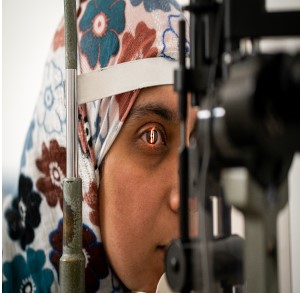
Diabetes affects 537 million people around the world; a number that is expected to increase to 643 million by 2030, according to International Diabetes Federation. Yet, diagnostic tools like blood sugar tests have their limitations and are not always accurate. Researchers from Hamad Medical Corporation and Hamad Bin Khalifa University have used data from 5,545 participants in Qatar to develop a deep learning model (DiaNet v2) that can diagnose diabetes with remarkable accuracy using retinal images.
The new model has achieved a 92% accuracy rate, compared to a rate of 84% in a previous model developed in 2021. Compared to other conventional diagnostic tools that rely on drawing blood samples to diagnose diabetes, the DiaNet v2 model is easier, faster, and cheaper, and doesn’t require people to fast.
“This non-invasive and accessible approach has the potential to revolutionize diabetes detection, especially in regions of the world with high rates of the disease, such as the Middle East and North Africa (MENA),” the researchers explain in a paper published in Scientific Reports.
Diabetes affects 73 million in the MENA region and is expected to rise to 634 million by 2030 and 136 million in 2045, according to International Diabetes Federation’s numbers in 2021.
“Our study aimed to enhance the accuracy of diabetes diagnosis by developing an improved predictive artificial intelligence model using retinal images from the Qatari population, and to address the limitations of current diagnostic methods. Early detection of the disease allows timely intervention,” says Tanvir Alam, assistant professor at the College of Science and Engineering at Hamad Bin Khalifa University in Qatar and lead author of the research.
Previous research has indicated that the retinas of people with diabetes normally show signs of damage from the disease; including small blood vessel damage, swelling, yellow deposits, bleeding, cloudy spots and areas with reduced blood flow. These signs make it possible to diagnose diabetes through retinal imaging. The side effects of diabetes on the retina, however, are usually a late complication of the disease, and difficult to diagnose early using retinal imaging.
Advancing and complementing diagnostics
Diabetes is diagnosed and controlled by measuring blood sugar levels. The glucose is measured randomly during the day, or after fasting for at least eight hours. It is also measured using the HbA1C test, which indicates the average blood glucose levels in the past three months. The oral
glucose tolerance test can also be performed to screen for type 2 diabetes, prediabetes and gestational diabetes and entails drawing a blood sample to measure glucose levels after fasting for at least eight hours, then redrawing to measure levels two hours later after drinking a glucose solution. These methods can be uncomfortable for some people and some can cause dizziness and nausea.
“Although [these methods] are widely used, these tests have limitations. For example, the fasting glucose test has less sensitivity in identifying true patients, leaving 30% of diabetes cases undiagnosed, while the oral glucose tolerance test is labor intensive, and misclassifies 12% of cases,” says Alam.
The DiaNet v2 model was trained on 16,000 retinal images taken from 52,540 people with diabetes and 3,005 healthy controls.
The model distinguished people with diabetes from the control group with a 91% specificity rate. There are other attempts at diagnosing diabetes using machine learning-based retinal images, including a research published in BMJ Open Diabetes Research & Care in 2022 that developed a model with a similar sensitivity rate of 92% and specificity rate of 96.2% using data from 2,221 participants from Hong Kong and the United Kingdom. The DiaNet model, however, is the first developed using regional data and with a larger pool of data.
Alm points out that previous research conducted by the team showed the usefulness of retinal images in early detection of cardiovascular diseases and the classification of various eye conditions, including diabetic retinopathy, diabetic macular edema, and glaucoma.
“This diagnostic approach is a rapid, non-invasive screening for diabetes, and reduces the cost of the disease and the burden on healthcare professionals while enhancing the quality of diagnosis and supporting decision-making during examination,” says Alm.
Gamal Al-Mashad, professor of ophthalmology at Zagazig University in Egypt, who was not involved in the research, says detecting the disease based on retinal images can be a complementary method that facilitates diagnosis in remote areas that do not have enough traditional diagnostic tools available.
The retinal imaging method does not replace traditional diagnostic methods, but can facilitate effective disease screening in areas with limited resources and areas that lack adequate healthcare infrastructure.
“By integrating our AI-powered solution with telemedicine platforms, patients in remote areas can connect seamlessly, enabling rapid and accurate diagnosis of the disease within minutes and at the lowest cost,” says Alm.
doi:10.1038/nmiddleeast.2024.80
River subbasins more depleted than official figures show
09 April 2024
Arab countries perform poorly on climate action scorecard
24 March 2024
12 March 2024
Sign-up to receive our e-alert update every two weeks to keep up with everything new on the portal.
Sign up for e-alerts
Stay connected: